┌────── ■Ultrasonic Imaging Laboratory■ ──────┐
|
|
Research Title: All optical based intravascular ultrasound/photoacoustic imaging: scanhead and system design and development |
|
|
PI: Professor Pai-Chi Li |
|
|
Co-PI: Dr. Lung-Chun Lin, NTU Hospital |
|
|
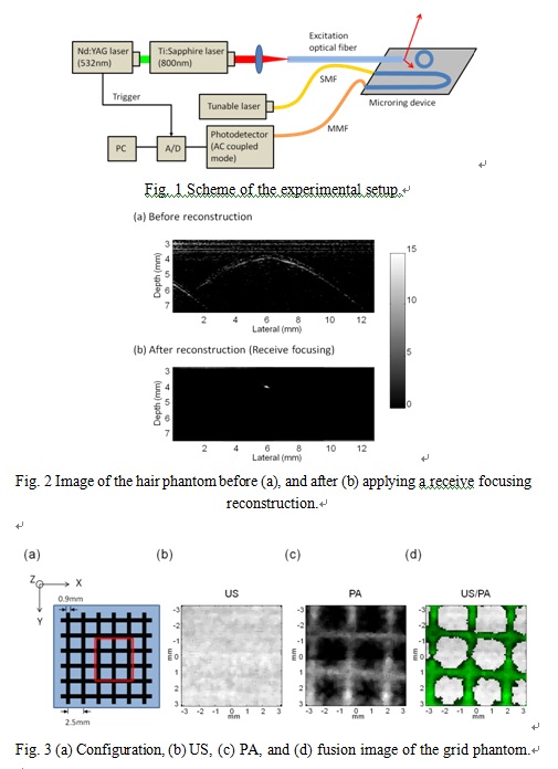
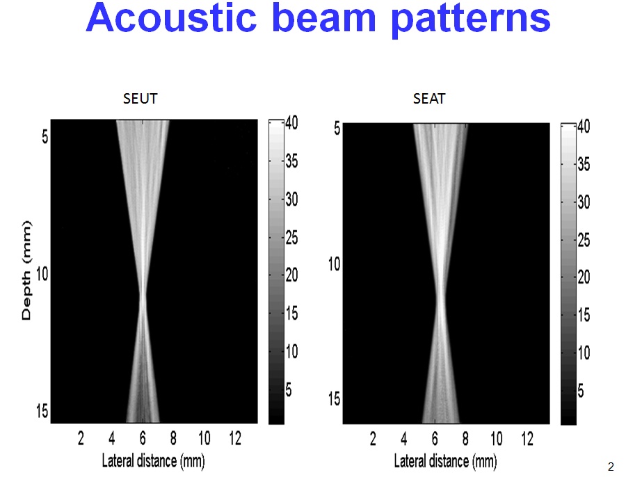
Abstract: Atherosclerotic diseases are a serious problem in modern counties, and it has been one of the major causes of acute ischemic heart diseases. For diagnosis, intravascular ultrasound has been a widely accepted tool. In order to further improve intravascular image quality and to provide a better diagnostic tool, the purpose of this project is to develop advanced scan head technologies for intravascular imaging. The clinical significance of this project is the combination of ultrasound and photoacoustics, so that more complete information can be provided. To this end, the scanhead at the catheter tip needs to have fundamentally new designs, so that it can be small size, high frame rate, low cost and high quality. To achieve this goal, the advanced technologies of this project include: - All optical based ultrasound and photoacoustic imaging technologies: Based on the photoacoustic response from a thin film and proper design of light reflection, the laser can be used to excite high frequency ultrasound to perform ultrasound imaging. At the same time, utilizing light reflection, the reflected light can also be used for photoacoustic imaging at the same time. On receive, using micro-ring resonators as the ultrasound sensor, the acoustic echoes from both ultrasound imaging and photoacoustic imaging can be simultaneously detected. - Subband imaging for improved contrast: Because the frequency content of the acoustic echoes from ultrasound scattering and photoacoustic generation is different, the proposed subband imaging method can be used to separate these two types of signals and an optimal contrast enhancement method by properly combining these two signals can be developed. - 360 degree transmit and receive method: With this method, only a single laser pulse is needed to acquire an ultrasound image frame and a photoacoustic image frame. By doing so, we can effectively reduce the requirement on laser pulse repetition frequency and thus the system complexity can be greatly reduced. - Real-time ultrasound and photoacoustic imaging: This is based on previous achievements of the research team. A real-time system is readily available to be combined with the new scanhead proposed in this project, and a complete real-time ultrasound and photoacoustic system for intravascular imaging can be produced. Ex vivo experiments will also be conducted to further verify feasibility and performance of the proposed methods. Based on our previous research results and with the successful execution of this project, we will be in the leading position in diagnostic technologies for intravascular imaging of atherosclerosis and make real clinical contributions in this area.
|
|
|
|
|
|
|
|
|
|
|
|
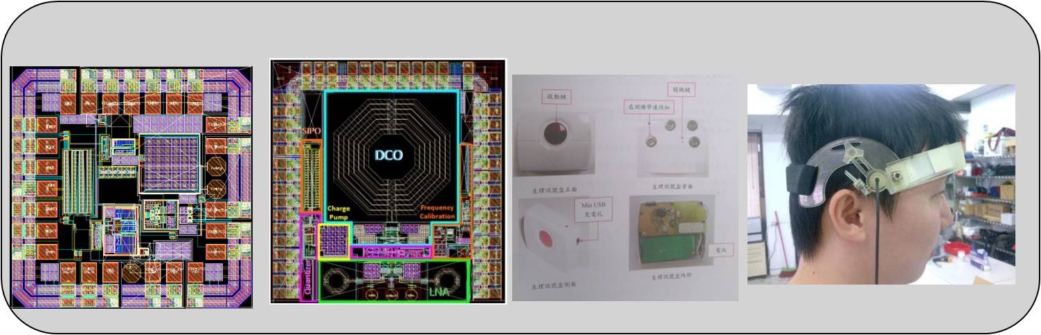
Research Title: Development of Advanced Biomedical ICs and Integration of Medical Systems |
|
|
PI: Professor Pai-Chi Li |
|
|
Co-PI: Professor Chen-Tung Yen, Professor An-Yu Wu, Professor Shen-Iuan Liu, Professor Tsung-Hsien Lin, Professor Jri Lee, National Taiwan University. Dr. Lung-Chun Lin, Dr. Dar-Ming Lai, NTU Hospital. |
|
|
Abstract: The long term goal of this integrated project is to develop next generation biomedical electronics and system integration technologies for biomedical applications. By combining low power wireless transmission, advanced adaptive imaging technologies for portable devices, high performance digital signal processing engine, GPU-based medical computing platform, wireless power transmission and 60 GHz wireless transmission, leading technologies will be developed and transferred to the industry for important biomedical applications. The research team of this project consists of outstanding researchers from disciplines of engineering, medicine and fundamental sciences. In addition, the following three system platforms will be used as vehicles for technology development: low power/wireless physiology monitors, portable adaptive ultrasonic imaging system and wireless neural stimulator. To achieve these goals, the project will consist of the following main items: - Low power wireless communication - Adaptive ultrasonic imaging methods on portable platforms - Ultrasonic wireless power transmission - Wireless neural stimulator - 60 GHz transmitters and receivers for imaging applications - Miniaturized piezoelectric sensors The success of this project will put the research team in the forefront position in the world, and assist the transformation of domestic IT industry to next generation biomedical electronics businesses. Sub-project 1
Sub-project 2
Sub-project 3
Sub-project 4 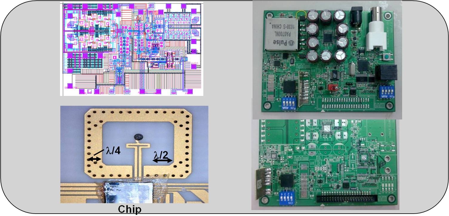
|
|
|
|
|
|
|
|
|
|
|
|
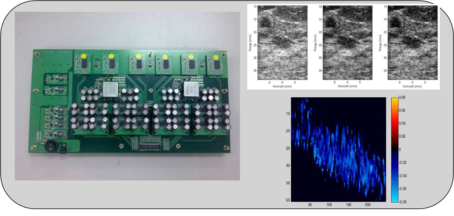
Research Title: Development of IVPA/IVUS imaging technologies and investigation on ultrasound-assisted thrombolysis |
|
|
PI: Professor Pai-Chi Li |
|
|
Co-PI: Dr. Lung-Chun Lin, NTU Hospital |
|
|
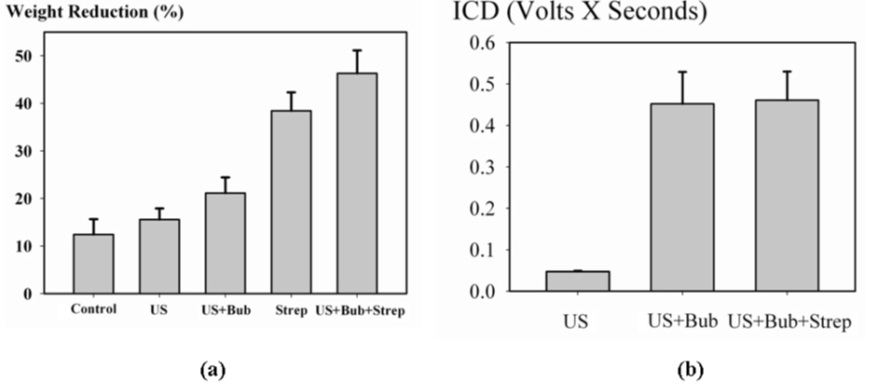
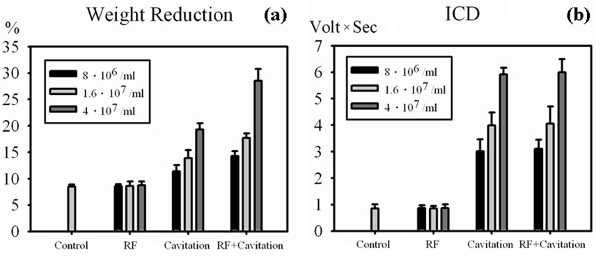
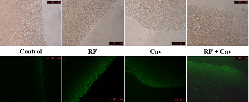
Abstract: The main goal of this three-year project is to develop advanced imaging and therapy technologies for various stages of vascular diseasess. Specifically, we will develop multi-modality imaging technologies for atherosclerotic plaque imaging and staging, and image guided thrombolysis technologies that is assisted by specific binding and acoustic cavitation effects. The multi-modality imaging will combine both intravascular photoacoustic imaging (IVPA) and intravascular ultrasonic imaging (IVUS). IVPA primarily shows information related to optical absorption, and IVUS is primarily based on acoustic scattering. Therefore, the combination can provide more clinical diagnostic information that cannot be provided by any other existing imaging technologies. In addition, correlation based ultrasonic flow estimation methods can also be incorporated in order to provide quantitative blood flow information. Because both modalities form images based on the detected acoustic waves, the multi-modality imaging system can be integrated effectively. On the other hand, other imaging methods, such as ultrasonic elasticity imaging for vessel characterization and plaque staging, can also be effectively integrated to provide additional diagnostic information in the future. The other major component of this project is on thrombolysis. In particular, ultrasound and microbubbles will be exploited in order to understand the potential of cavitation-assisted thrombolysis. Moreover, molecular probes (conjugated microbubbles) will be developed so that these probes can target thrombus, and it is expected that this can further enhance the thrombolysis effectiveness. In order to achieve these goals, we will also work with National Taiwan University Hospital on the animal models for atherosclerosis. We will work with our international collaborator at the University of New Mexico as well, on a novel CMUT based photoacoustic transducer. The development and the applications of the CMUT transducer are also a pioneering work in the field. To this end, the specific aims of this project include: - Development of a prototype photoacoustic probe - Investigation of cavitation assisted thrombolysis - Development of ultrasonic molecular probes for thrombus targeting - Investigation of targeted cavitation assisted thrombolysis - IVPA/IVUS plaque characterization - Development of dual mode IVPA/IVUS imaging system - Image guided thrombolysis Success of this project will put the research team in a leading position in the world in the area of intravascular imaging and enhanced thrombolysis
|
|
|
|
|
|
|
|
|
|
|
|
Research Title: Quantitative study of US based targeted therapy: the use of US/PET and US/MRI molecular probes |
|
|
PI: Professor Pai-Chi Li |
|
|
Co-PI: Dr. Kai-Yuan Tzen, NTU Hospital, Professor Hsin-Ell Wang, National Yang-Ming University |
|
|
Abstract: Targeted drug delivery has gained wide interest as a more effective therapy method. In addition, ultrasound has been proven to be able to enhance the drug delivery effectiveness and it is possible to be combined with ultrasonic molecular imaging for image guiding and evaluation during therapy. However, one limitation of ultrasound is that it is not capable of performing whole-body scanning. Thus, it cannot be used for quantitative monitoring of whole-body biodistribution, circulation and metabolism. Without such important information as metabolism, distribution of drug accumulation, treatment effects and damage in surrounding tissue, successful treatment planning will not be possible. Thus, it is the purpose of this study to develop technologies for quantitative whole-body evaluation of targeted drug delivery and cancer therapy effectiveness by using multi-modality molecular imaging. In this project, breast cancer MDA-MB-231 will be used as the primary cell line and disease model, with Paclitaxel being the main drug. To this end, the project has the following specific aims: - Preparation and production of multi-modality molecular probes. - Preparation and production of VEGFR2 18F-SFB-MBs and Albumin-(Gd-DTPA)-MBs. - Preparation and production of VEGFR2 microbubbles. - Targeted drug delivery and ultrasonic molecular imaging. - Production and analysis of targeting drug-carrying microbubbles. - High frequency small animal ultrasonic imaging - Multi-modality imaging and targeted drug delivery - Quantitative US/PET small animal imaging - Image registration for US/PET imaging and US/MRI imaging. Success of this project will put the team in a leading position in multi-modality molecular imaging. It will also contribute to drug delivery and targeted therapy, with the main goal of quantitative evaluation of targeted drug release and cancer treatment.
|
|
|
|
|
|
|
|
|
|
|
|
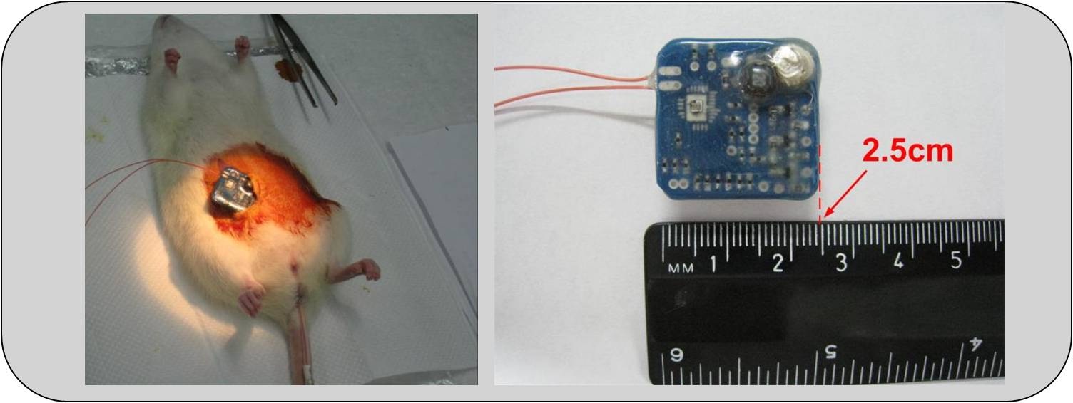
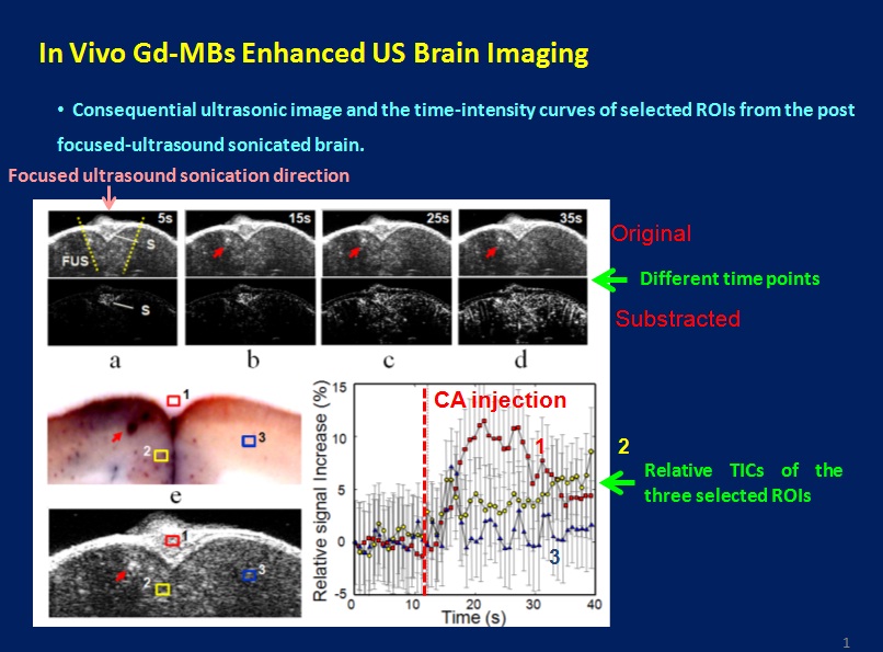
Research Title: Multi-modality/mutli-targeting micro-imaging for preclinical research |
|
|
PI: Professor Pai-Chi Li |
|
|
Co-PI (Foresight Taiwan): Professor Ji-Wen Sun, Academia Sinica |
|
|
Abstract: This is a continuation project, with the main goal being to target to the pre-clinical small animal imaging market, which will grow to a size of USD 556M in 2012 in the US alone. Success of our previous research on imaging system design, device development and temperature monitoring will be transformed into an effective tool needed in this emerging market of drug development and cancer research. To this end, specific aims of this project include: - Integration of a real-time, 20MHz, ultrasonic imaging/photoacoustic imaging/temperature monitoring system. - Array-based high frame rate photoacoustic and ultrasonic imaging methods. - Quantitative temperature monitoring for targeted photothermal therapy. - Modulation methods of CW diode laser for effective photoacoustic generation. With the continuing success of this project, it will lead to opportunities for Taiwan to become a major player in this emerging market.
|
|
|
|
|
|
|
|
|
|